
No Activation, No Strength: A Practical Approach to AMI After ACL Reconstruction
Arthrogenic muscle inhibition (AMI) is a neurological barrier that halts early rehabilitation in its tracks. To restore true function, clinicians must look beyond traditional mechanical loading and prioritize targeted, neuro-centric strategies that unlock the nervous system.
July 8, 2026
8 min. read
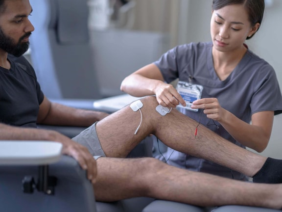
A patient presents six weeks after anterior cruciate ligament (ACL) reconstruction with full range of motion (ROM), minimal pain, and no significant swelling. Yet, they look up from the table with a frustrating question: “Why can't I activate my quads?”
This scenario is all too common in orthopedic rehabilitation. Clinicians frequently encounter patients who check every structural box for recovery but still struggle with a profound disconnect between their brain and their knee. To truly help these patients overcome this invisible barrier, we must look beyond traditional mechanical loading and target the underlying neurological dysfunction that halts early rehabilitation in its tracks.
What is really happening?
ACL reconstruction is one of the most common sports-related procedures. However, it is frequently associated with long-term functional deficits when neural inhibition is not properly addressed in the early stages of rehabilitation.
One of the primary roadblocks during recovery is arthrogenic muscle inhibition (AMI), a neural mechanism that limits the ability to fully contract the quadriceps muscle, particularly the vastus medialis.1 AMI is highly prevalent following ACL reconstruction and can persist throughout the entire rehabilitation process, with reported incidence rates of up to 56 percent at six weeks post-injury.2
For this reason, addressing AMI early is critical. Failure to do so may lead to suboptimal recovery, persistent strength deficits, and long-term functional limitations.
From a pathophysiological perspective, AMI is not just a localized joint issue; rather, it involves both the peripheral and central nervous systems. Following reconstruction, a cascade of events, including inflammation, joint effusion, and altered mechanoreceptor signaling, modifies afferent (sensory) input. This increases neural inhibition and reduces the activation capacity of the quadriceps extensor muscles. Furthermore, cortical changes alter motor control, which perpetuates the muscle inhibition loop.
Ultimately, this is not just a problem of muscle strength, but a fundamental issue of neuromuscular activation.3
Why traditional rehab fails
When a patient comes in for their first consultation after surgery, I always explain a vital concept: Traditional ACL rehabilitation protocols often focus on restoring strength as quickly as possible. However, without proper quadriceps activation, any attempt to build strength will be limited.
I like to tell my patients, “It makes no sense to talk about strength if the muscle is not activating properly. It’s like trying to drive a car at 100 kilometers per hour when it won’t even start.”
This is where the real problem lies. Traditional protocols assume that progressively increasing load, volume, and exercise progression will naturally restore muscle function. But when AMI is present, the limiting factor is neural connectivity. The muscle tissue is ready, but the nervous system is not.
As a result, patients may appear to improve their strength on paper without actually achieving true quadriceps activation. Instead, they develop compensatory movement strategies, particularly at the hip, to bypass knee extension. This explains why many patients diligently follow rehabilitation protocols yet still fail to fully restore quadriceps neuromuscular function. Until neural inhibition is systematically addressed, adding more strength work is counterproductive.
Major clinical consequences
When AMI is left unaddressed in the early stages following ACL reconstruction, it triggers a domino effect of clinical consequences that can compromise long-term outcomes.2
Consequence | Clinical impact |
Persistent muscle weakness and atrophy | Impaired motor unit recruitment leads to chronic quadriceps weakness, particularly in the vastus medialis. |
Joint stiffness and extension deficit | Poor activation can result in a “pseudolocked” knee, contributing to stiffness, extension deficits, and potential cyclops lesion development. |
Altered gait and mobility | Compensatory movement patterns reduce gait efficiency and accelerate fatigue during daily activities. |
Delayed rehabilitation progression | AMI limits effective participation in early rehabilitation, stalling recovery timelines and delaying return-to-sport testing. |
Long-term degenerative changes | Chronic deficits in muscle function and joint loading alter biomechanics, potentially contributing to cartilage degeneration and early osteoarthritis. |
Increased reinjury risk | Deficits in neuromuscular control and joint stability increase the likelihood of subsequent knee injuries. |
Practical solutions: A phase-by-phase approach
Clinical case example: A 20-year-old patient presents 10 days post-ACL reconstruction with moderate joint effusion, pain (VAS 5/10), an inability to voluntarily contract the quadriceps, a 10-degree knee extension deficit, and clear clinical signs of AMI.
Weeks zero to four: "Unlock the system" and restore activation
The primary goals for this initial phase include:
Reduce pain and joint effusion
Decrease neural inhibition
Restore voluntary quadriceps activation
Achieve full knee extension
At this stage, rehabilitation should not focus on hypertrophy or strength development, but rather on restoring central neural drive.
The intervention strategy should be implemented via structured daily rehabilitation, Monday through Friday, utilizing the following modalities:
1. Structured exercise (the foundation of treatment)
Structured exercise should be implemented as the primary intervention from the earliest postoperative days. The goal is to restore neural drive to the quadriceps using specific principles:
Progression: Carefully adapted to the patient's pain, inflammation, and motor control.
Load: Just enough to elicit activation, even at low intensity.
Neural intention: A strict emphasis on maximal voluntary contraction.
As a practical clinical cue, I commonly tell the patient: “Push the back of your knee down into the table and try to lift your kneecap upward.”
Physiologically, early exercise acts as a neural primer by increasing motor system excitability, reducing arthrogenic inhibition, and improving voluntary quadriceps activation.
2. Cryotherapy (pre-activation strategy)
Apply cryotherapy for 15 minutes immediately prior to each session. Cold therapy reduces inhibitory afferent input from the swollen joint, which facilitates motor neuron excitability and makes it easier for the patient to voluntarily contract the quadriceps.4
Educating the patient on how cold therapy alters nerve signaling and aids recovery is crucial for compliance; the patient education video below serves as an excellent resource to help them understand the science behind cryotherapy.
3. Motor imagery (cortical priming)
Performed before active exercise (three sets of 10 repetitions). Instruct the patient to vividly imagine a strong knee extension and a powerful quadriceps contraction. This cognitive strategy increases cortical activity, enhances corticospinal excitability, and reduces central inhibition to prime subsequent motor output.
4. Biofeedback
To improve muscle recruitment and motor control, begin by placing visual or electromyographic (EMG) biofeedback on the contralateral (unoperated) limb. Once the patient understands the target contraction, transfer the feedback to the operated limb (three sets of 15 repetitions per limb). This cross-education approach increases corticospinal excitability, reduces interhemispheric inhibition, and accelerates motor learning.5
Weeks two to four: Consolidate activation and introduce load
The primary goals for this progression phase include:
Achieve consistent, reliable quadriceps activation
Eliminate compensatory movement patterns
Initiate progressive loading
The intervention strategy during this phase shifts toward progressive load introduction, provided neuromuscular activation is sustained:
1. Maintain activation
Progress to loaded isometric exercises while maintaining a strict emphasis on motor control quality.
2. Introduce open kinetic chain (OKC) exercises
Implement progressive, non-weight-bearing knee extensions. These are essential for isolating and restoring specific quadriceps function.
3. Basic closed kinetic chain (CKC) exercises
Introduce controlled, early weight-bearing movements such as mini-squats and step-ups.
Clinical consideration: If adequate quadriceps activation is not achieved by this stage, the clinician should pause progression, reassess earlier-phase interventions, and reinforce neural de-inhibition strategies.
Conclusion: The neurological priority
In the early phases following ACL reconstruction, the primary limiting factor is rarely a lack of muscle capacity—it is the presence of neural inhibition. When a patient asks why they cannot fire their quadriceps six weeks post-op, adding more weight or forcing more repetitions will not bypass the block. Until the neural pathways are cleared, the muscle tissue simply cannot receive the signal to adapt.
To optimize long-term clinical outcomes, rehabilitation must follow a logical, neuro-centric progression sequence rather than a simple timeline of tissue healing:
Reduce inhibition
Restore activation
Facilitate activation if necessary
Build strength
Transfer to function
By shifting your focus from mechanical strength to neuromuscular activation during the critical first four weeks, you effectively “unlock the system.” Implementing targeted strategies like pre-activity cryotherapy, biofeedback cross-education, and cortical priming ensures that the nervous system is primed. When you finally do transition to traditional loading and functional progression, your patient will have the neuromuscular foundation required to build true strength.
Remember: No activation, no adaptation. Target AMI first to clear a predictable path for a safe, successful return to sport.
References
Hopkins, J. T., & Ingersoll, C. D. (2022). Arthrogenic muscle inhibition: 20 years on. Journal of Sport Rehabilitation, 31(6), 665–666. https://pubmed.ncbi.nlm.nih.gov/35894917/
Sonnery-Cottet, B., Hopper, G. P., Gousopoulos, L., Vieira, T. D., Thaunat, M., Fayard, J. M., Freychet, B., Ouanezar, H., Cavaignac, E., & Saithna, A. (2022). Arthrogenic muscle inhibition following knee injury or surgery: Pathophysiology, classification, and treatment. Video Journal of Sports Medicine, 2(3), 26350254221086295. https://pubmed.ncbi.nlm.nih.gov/40308858/
Paço, M., Peysson, M., Dumont, E., Correia, M., Quialheiro, A., & Chaves, P. (2024). The effect of physiotherapy on arthrogenic muscle inhibition after ACL injury or reconstruction: A systematic review. Life, 14(12), 1586. https://pubmed.ncbi.nlm.nih.gov/39768294/
Rice, D., McNair, P. J., & Dalbeth, N. (2009). Effects of cryotherapy on arthrogenic muscle inhibition using an experimental model of knee swelling. Arthritis & Rheumatism, 61(1), 78–83. https://pubmed.ncbi.nlm.nih.gov/19116960/
Richaud, T., Lacaze, K., Fassio, A., Nicolas, P., Ourliac, M., Hennart, B., Ina, J. G., Sonnery-Cottet, B., & Cavaignac, E. (2024). How biofeedback with surface EMG can contribute to the diagnosis and treatment of AMI in the knee. Video Journal of Sports Medicine, 4(4), 26350254241241084. https://pmc.ncbi.nlm.nih.gov/articles/PMC11752188/
Meet the Author


Subscribe to Our Newsletter
Related Posts

September 9, 2024
Top 10 Essential Strategies for ACL Rehabilitation Success
By Kyle Matsel PT and Phil Plisky

December 20, 2024
Effective Meniscus Injury Exercises for Rehabilitation and Recovery
By Medbridge

November 15, 2024
Top Exercises for the Early Stages of Knee Pain Recovery
By Medbridge
July 11, 2016
When to Return to Sport? A Brief History of ACL Rehabilitation Approaches
By Terry Malone