Musculoskeletal Radiology and Imaging: Fundamentals, Part 2
Presented by Dr. Robert Boyles
This course expands on foundational principles and explores advanced musculoskeletal imaging modalities to help clinicians enhance diagnostic decision-making and optimize patient care. Clinicians will examine the indications, benefits, and limitations of fluoroscopy, nuclear medicine, bone scans, ultrasound imaging, computed tomography (CT), and magnetic resonance imaging (MRI). The course analyzes the imaging mechanics, clinical relevance, and appropriate application of each modality in musculoskeletal pathology. Physical therapists, athletic trainers, physician assistants, and other healthcare professionals will learn to select suitable imaging techniques for diverse patient presentations in outpatient, surgical, and acute care settings.
Learning Objectives
- Build on your basic knowledge of musculoskeletal imaging modalities by identifying the clinical purpose and typical use cases for X-ray, MRI, CT, and ultrasound
- Recognize when musculoskeletal imaging is appropriate by matching common clinical presentations with the most relevant imaging modality
- Identify key properties and limitations of commonly used imaging modalities in detecting specific musculoskeletal pathologies (e.g., fractures, soft tissue injuries, degenerative changes)
- Differentiate imaging modalities based on contrast resolution, safety, accessibility, and diagnostic utility
- Visualize how imaging findings integrate into the clinical examination to support clinical reasoning, referral decisions, and interdisciplinary communication
- Relate imaging findings to occupational performance by identifying how musculoskeletal conditions seen on imaging may impact ADLs, IADLs, and functional participation in daily roles
Meet your instructor

Dr. Robert Boyles
Dr. Robert Boyles is a clinical professor and former program director of the University of Puget Sound’s Doctor of Physical Therapy program in Tacoma, Washington. Previously, he was on faculty as associate professor and director of clinical education of the US Army-Baylor University DPT program. His primary areas of…
Chapters & learning objectives
1. Fluoroscopy and Nuclear Imaging
This chapter introduces fluoroscopy and nuclear imaging, focusing on their unique ability to provide real-time imaging or physiologic information. Fluoroscopy enables dynamic imaging during procedures such as fracture fixation and arthrography, while nuclear imaging highlights metabolic activity and provides enhanced imaging to detect pathology, such as infection or tumor involvement. Understanding these tools is essential for guiding interventions and identifying changes not seen with standard radiography.

2. Bone Scan
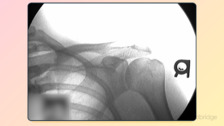
This chapter examines bone scintigraphy, a sensitive imaging modality that detects increased bone metabolic activity. Clinicians will learn how bone scans aid in the early detection of stress fractures, avascular necrosis, infections, and metastatic disease. The utility of the three-phase bone scan, particularly for subtle injuries such as nondisplaced scaphoid fractures, and metabolic activity in bone to detect stress reactions and tumors, is highlighted to support more accurate and timely diagnoses.

3. Ultrasound
Focusing on musculoskeletal ultrasound, this chapter explores its dynamic imaging capabilities for soft tissue evaluation, including tendons, ligaments, and fluid collections. Clinicians will understand the advantages of ultrasound—such as portability, low cost, and absence of radiation—alongside its limitations, including operator dependency and reduced bone penetration. Real-world applications in evaluating rotator cuff tears, nerve entrapment, and healing fractures are emphasized.

4. Computed Tomography (CT)
This chapter covers CT imaging, detailing its use in evaluating complex fractures, bone alignment, and degenerative changes. With fast acquisition and high-resolution 3D reconstructions, CT is often the modality of choice in acute trauma. Limitations, such as high radiation exposure and reduced soft-tissue contrast, are discussed to guide safe and appropriate use in musculoskeletal care.

5. Magnetic Resonance Imaging (MRI)
In this chapter, learners will explore MRI as the gold standard for evaluating soft tissue pathology, including ligament tears, bone marrow changes, and occult fractures. The chapter outlines MRI sequences, safety considerations, and the advantages of contrast resolution in distinguishing between normal and pathologic tissue. By understanding MRI’s strengths and contraindications, clinicians can better determine when this advanced imaging is warranted.
Frequently asked questions
Everything you need to know about Medbridge courses.
Who creates Medbridge courses?
We work with industry-leaders, top researchers, and consultants to build content roadmaps that are then structured into courses, filmed, and edited by our production team before being launched to our site.
How often does Medbridge release new courses?
New courses are added monthly and are automatically included in your subscription as they launch.
How often does Medbridge update courses?
Medbridge reviews its courses annually for relevance and to assess if content is up to date. Based on these reviews it may be determined that a course is out of date resulting in the course being re-filmed or retired, if the content is no longer needed (e.g. a replacement course already exists, the concepts are no longer best practice, etc.).
How many courses does Medbridge offer?
We have over 3,000 accredited courses and we are continually updating our library with new courses. Check our course library for the most up-to-date count for your discipline.
Are there any additional fees for taking a Medbridge course?
There are no additional fees for taking a Medbridge course, obtaining a course certificate of completion, earning CEUs as a subscriber, or accessing any of the additional tools your subscription may include.
Is there a limit to the number of courses I can take?
There is no limit to the number of courses you can take as a subscriber! As a subscriber to Medbridge, you have unlimited access to over 3,000+ accredited CE courses.
If you are a Premium subscriber, you also have unlimited access to our Patient Engagement Tools such as the Home Exercise Program, Patient Education Library, Orthopedic Exam Tests, and Manual Therapy Techniques. We have over 7,000 exercises and over 650+ videos and handouts of patient education resources with more exercises and patient education added to the library based on subscriber feedback, volume of request and specialities.
What is your refund policy?
You are eligible for a refund provided your request is received within 30 days of your subscription purchase and your account has no activity.