Beyond Standard Protocols: A Practical Approach to Optimizing Shoulder Arthroplasty Outcomes
Move beyond generic checklists and elevate your clinical practice with evidence-based strategies designed to optimize recovery for the modern shoulder arthroplasty patient. Master short-lever facilitation and kinetic chain releases to bridge the gap between standard protocols and superior functional outcomes.
April 24, 2026
11 min. read
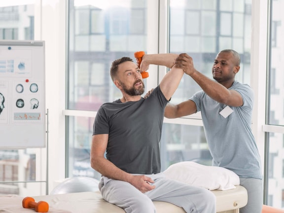
When a total shoulder arthroplasty (TSA) patient shows up on my schedule four days post-op, I don't just reach for a generic PDF protocol. If we’re being honest, many of those protocols are a mindless collection of ball squeezes and pendulums that fail to address the complexity of the modern surgical patient.
Instead of falling back on autopilot, I recognize that effective rehabilitation begins with establishing a partnership. I start by opening the floor to their questions. These are high-expectation consumers who have often spent years in pain, and they need to know that we aren't just going through the motions. We are engaging in high-level manipulative therapy and science-based facilitation.
The surgical landscape has shifted from historical six-week hospital stays and blood transfusions to modern same-day outpatient centers. If our rehab doesn't continue to evolve and innovate at a similar pace, we are failing to practice at the highest level of our license.
The reverse revolution: why 80 percent?
Initially, reverse total shoulder arthroplasty (rTSA) was a niche procedure reserved for patients with rotator cuff deficiency. Today, it accounts for roughly 80 percent of all shoulder replacements.1
Surgeons are moving toward rTSA even with an intact cuff because the clinical results are incredibly consistent. We know the natural history: roughly 60 percent of patients over age 60 have asymptomatic tears.2 By choosing rTSA, we bypass the risk of a "cuff blowout" later, ensuring a prosthesis with a 75 percent survival rate at 20 years.3,4 In an anatomic shoulder (aTSA), we are reliant on a thinning, poorly vascularized rotator cuff to center the ball; in rTSA, we utilize the mechanical advantage of the deltoid.
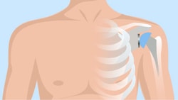
Understanding the anterosuperior escape
To appreciate why we train the way we do, we have to understand the pathology of the arthritic shoulder. In a healthy joint, the cuff produces an inferior-posterior glide to keep the humeral head centered. When the cuff is lousy, we see anterosuperior humeral escape—a pseudo-paresis where the ball migrates up and in against the glenoid fossa.
When you see this in the clinic, you realize that traditional long-lever exercises in early phases only worsen this migration. We must optimize the center of rotation before we ever ask for active elevation.
The subscapularis: tendon vs. bone
A critical clinical gem is knowing exactly how the surgeon accessed the joint. Traditionally, they detached the subscapularis tendon and reattached it (a tendon-to-bone repair with notoriously poor blood supply).
However, many modern surgeons perform a lesser tuberosity osteotomy (LTO). They shave a piece of bone off with the tendon attached, and then fix it back bone-to-bone. Because bone heals significantly better than tendon, this gives us the confidence to be slightly more assertive in our progressions, though we still respect the standard 30-degree external rotation limit for the first four weeks to protect that repair.
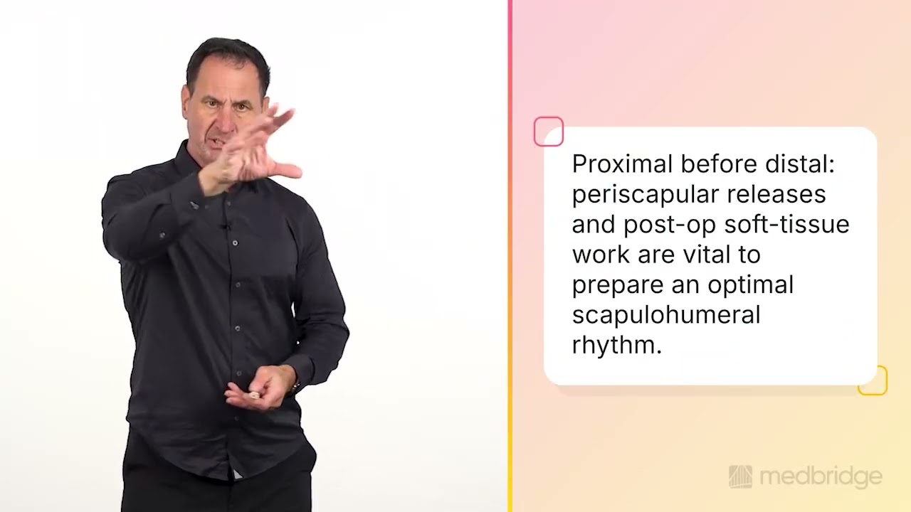
The day four framework: proximal before distal
In the first 21 days, my focus is on the core triad: proximal stability, distal mobility, and early facilitation.
Thoracic extension (proximal stability): Everyone can do this, and it is vital for optimal scapulohumeral rhythm. By addressing the thoracic spine early, we prevent the shrugging compensations that often occur when a patient lacks the postural base to support humeral elevation.
"See your elbow" (distal mobility and protection): This is my non-negotiable rule for the first six to eight weeks. If the patient cannot see their elbow, they are likely in a position of abduction and external rotation that places excessive stress on the anterior capsule and risks dislocation. By keeping the arm in the plane of the scapula, where they can clearly see the elbow, we protect the repair while maintaining distal hand and wrist function.
Neuromuscular electrical stimulation (early facilitation): Atrophy doesn't wait for your six-week follow-up. For every day we don't work the deltoid, we lose three to four days of functional recovery later. If there are no contraindications, I use neuromuscular electrical stimulation (NMES) on the anterior and middle deltoid fibers immediately to combat atrophy and provide involuntary motor fiber recruitment.
Rethinking the prime mover: short-lever facilitation
In a reverse shoulder, the deltoid is no longer just an elevator. It’s also a humeral head compressor. Because the cuff is gone, we use the short-lever concept to train the deltoid without over-stressing the joint.
The palmar grasp reflex: Traditional ball squeezes are just circulatory exercises. But if you have the patient squeeze the ball and flex the bicep simultaneously, you’ll feel the deltoid kick in with an involuntary isometric contraction.
The thumb cross technique: Palms together, thumbs crossed, fingertips under the chin. Raising only from 70 to 100 degrees facilitates the anterior and middle heads while preventing the neck (levator/upper trap) from taking over the movement.
Manual therapy and the kinetic chain
Patients often complain that their neck is "killing them" post-op. This is usually due to pre-operative guarding and over-activation of the levator scapula.
Levator pin and stretch: Apply pressure to the origin at the spine of the scapula and have the patient look into their opposite armpit. It’s a game-changer that prepares them for smoother active motion.5
Pec minor release: A tight pec minor tips the scapula anteriorly, altering the center of rotation and killing your external rotation gains. Rotate it back manually while having the patient engage in diaphragmatic breathing. We must treat these patients systemically. Their myofascial pain isn't just "bad luck"—it's a breathing and postural compensation.5
The hip drop method
The latissimus dorsi connects to the gluteus maximus via the posterior oblique fascial plane. We can use the lower body to stretch the upper body.
The technique: When you hit a tissue wall in passive flexion, have the patient drop their knees to the opposite side. This immediately releases the shoulder by another 5 to 10 degrees through the fascial connection, bypassing patient apprehension without forcing the joint.
Range of motion expectations: anatomic vs. reverse
One of the most frequent questions patients ask is, "How much movement will I actually get back?" It is vital to set realistic expectations early, as the benchmarks differ based on the type of prosthesis used.
Average forward flexion
Anatomic TSA: Patients typically reach an average of 149 degrees. Most clinical outcomes fall within a range of 136 to 162 degrees.6
Reverse rTSA: Patients typically reach an average of 142 degrees. The standard clinical range is between 127 and 157 degrees.6
Average external rotation
Anatomic TSA: The average recovery for external rotation is 63 degrees.6
Reverse rTSA: The average is slightly lower at 57 degrees.6
Functional reach (behind the back to level L3)
Reaching the L3 level of the spine is often a challenge for the reverse shoulder patient due to the mechanical design of the implant.
Anatomic TSA: Approximately 68.7 percent of patients are able to reach this milestone.6
Reverse rTSA: Only 37.3 percent of patients reach this level of function.6
Return to high-level activity and sport
Standardized protocols are notoriously inconsistent regarding return-to-sport timelines, with reported rates ranging from 49 percent to as high as 96 percent. Only about 40 to 55 percent of patients report improved sporting outcomes compared to their pre-operative state, making it vital to manage expectations early.3
My clinical experience and the research suggest that while four to six months is the standard window for daily activities and significant strength, more strenuous activities like golf, swimming, or heavy weightlifting typically require seven to nine months to ensure long-term tissue integrity.
A helpful way to frame this for your patients is the 6-6-6 rule:
Six weeks to achieve initial motion
Six weeks to establish a strength base
Six months (total) before they can truly say they are back and would have the surgery again
When clearing patients for high-impact or overhead sports, look for functional milestones rather than just the date on the calendar. For example, golfers can often begin with short irons and mid-irons around the four- to six-month mark, but only after demonstrating sufficient scapular control and strength.
For high-demand patients, such as the cowboy returning to lassoing or the heavy lifter, we must facilitate the posterior and middle deltoid to the highest degree. In a reverse shoulder, these fibers now perform the mechanical work of the absent rotator cuff. I strictly advise against movements that dislocate the joint under load, such as dips, lat pull-downs, and wide-grip bench presses.
Ultimately, I tell my patients: "It’s your shoulder and your discharge. You can do what you want, but if you go against this rationale, you’re doing it against medical advice." We are playing the long game to ensure this prosthesis lasts 20 years, not just 20 months.
Final thoughts: the impairment-based clinician
Don't be a protocol zombie. Every patient follows the "Goldilocks" rule: some are too tight, others too loose, and a few are just right. If a patient is a "scar tissue match-rater" with a history of post-surgical stiffness, start moving them early; if they are hypermobile, prioritize protection. By integrating manual releases, kinetic chain stretches, and deltoid facilitation, we don’t just meet normative data, but we exceed it.
Achieving these results requires moving beyond checklists to treat the specific impairments on your table. I invite you to explore my Medbridge course series to further refine your clinical decision-making:
Optimizing Function After Shoulder Arthroplasty: Master rehab strategies for the surge in anatomic and reverse replacements, focusing on deltoid facilitation.
Optimizing Outcomes After Total Joint Arthroplasty: Shift from “mindless rehab” to high-level gait facilitation and clinical decision-making.
Optimizing Gait After Hip Arthroplasty: Achieve superior functional outcomes through evidence-based gait analysis and corrective exercise.
Optimizing Recovery After Knee Arthroplasty: Master interventions for quadriceps facilitation and terminal joint extension to bridge the satisfaction gap.
Optimizing Complex Cases in Joint Arthroplasty: Master troubleshooting frameworks and advanced motor control to resolve chronic pain and mechanical dysfunction.
By practicing at the highest level of your license, you ensure your patients don’t just get by but return to the high-level activities that define their quality of life.
References
Rees, A. B., Graham, G. D., Burger, J. M., Saltzman, B. M., Schiffern, S., Connor, P., & Hamid, N. (2025). Is formal physical therapy necessary after reverse total shoulder arthroplasty? A single-blinded, randomized controlled trial. JSES International, 9(4), 1232–1236. https://pubmed.ncbi.nlm.nih.gov/40959011/
Haddad, D. J., Rizvi, O. H., Sherman, N. C., & Hamilton, A. R. (2024). Reverse and anatomic shoulder arthroplasty regional usage and open payment analysis using the Centers for Medicare and Medicaid Services database. Journal of Shoulder and Elbow Arthroplasty, 8, 24715492231207278. https://pmc.ncbi.nlm.nih.gov/articles/PMC10860377/
Beleckas, C., Schodlbauer, D., Mousad, A., et al. (2025). Evaluation of new normal after shoulder arthroplasty: Comparison of anatomic vs. reverse total shoulder arthroplasty. Journal of Shoulder and Elbow Surgery, 34, S43–S49. https://www.jshoulderelbow.org/article/S1058-2746(25)00196-X/fulltext
Kirsch, J. M., Puzzitiello, R. N., Swanson, D., Le, K., Hart, P. A., Churchill, R., Elhassan, B., Warner, J. J. P., & Jawa, A. (2022). Outcomes after anatomic and reverse shoulder arthroplasty for the treatment of glenohumeral osteoarthritis: A propensity score-matched analysis. The Journal of Bone and Joint Surgery. American Volume, 104(15), 1362–1369. https://pubmed.ncbi.nlm.nih.gov/35867705/
Schick, S., Dombrowsky, A., Egbaria, J., Paul, K. D., Brabston, E., Momaya, A., & Ponce, B. (2023). Variability in physical therapy protocols following total shoulder arthroplasty. Clinics in Shoulder and Elbow, 26(3), 267–275. https://pubmed.ncbi.nlm.nih.gov/37559522/
Levy, O., Mullett, H., Roberts, S., & Copeland, S. (2008). The role of anterior deltoid reeducation in patients with massive irreparable degenerative rotator cuff tears. Journal of Shoulder and Elbow Surgery, 17(6), 863–870. https://pubmed.ncbi.nlm.nih.gov/18718765/
Below, watch John O’Halloran discuss his approach to total shoulder arthroplasty in this brief clip from his Medbridge course "Optimizing Function After Shoulder Arthroplasty."
Meet the Author

Subscribe to Our Newsletter
Related Posts

October 2, 2025
Evidence-Based Management of Upper Extremity Osteoarthritis: From Evaluation to Arthroplasty Rehab
By Christopher Bise

June 2, 2021
Shoulder Replacement: Addressing Shoulder Hike and Shrug
By John O'Halloran
July 19, 2016
Shoulder Replacement: How Can We Speed Up Recovery?
By John O'Halloran
August 16, 2017
Rotator Cuff Injury Assessment & Suggested HEP
By Lenny Macrina