
Chronic Ankle Instability: What You Might Be Missing
Chronic ankle instability is rarely just a ligament problem. Jared Vagy, PT, DPT, OCS, CSCS, shows how to assess the ankle, foot, and hip together and turn those findings into a practical rehabilitation progression.
October 16, 2026
8 min. read
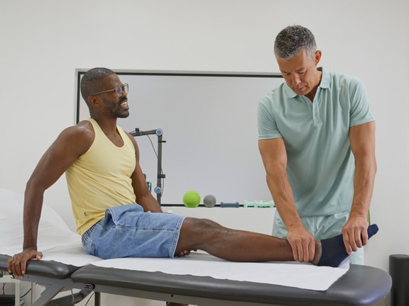
A patient tells you, “I sprained my ankle two years ago. It mostly healed, but it still feels unstable on uneven ground.” It’s tempting to blame the problem on loose ligaments and focus on ankle strengthening and balance exercises, and while those interventions may help, loose ligaments rarely explain the whole picture.
Chronic ankle instability can reflect a combination of ligament injury, mobility restrictions elsewhere in the ankle and foot, changes in muscle performance, and a movement strategy that does not distribute load well. The assessment should identify what keeps the ankle from feeling reliable during ordinary tasks such as walking on uneven ground, going down stairs, or stepping off a curb.
How to Assess Chronic Ankle Instability
Many clinicians begin with the standard anterior drawer test to determine whether the anterior talofibular ligament (ATFL) is affected. It is familiar and quick. A positive anterior drawer test can support ATFL involvement, but a negative result does not rule it out, particularly early after an injury.
When chronic ATFL involvement is suspected, the reverse anterolateral drawer test can supplement the examination. In one diagnostic study of chronic ATFL injury, the reverse test demonstrated 89.4 percent sensitivity, 89.7 percent specificity, and 89.6 percent overall accuracy. In the same comparison, the standard anterior drawer test had 22.4 percent sensitivity and the anterolateral drawer test had 47.3 percent sensitivity.
Interpret the result alongside a history of recurrent giving way, tenderness over the ATFL, and the person’s functional complaints. A positive reverse anterolateral drawer test can strengthen the case that residual ligament insufficiency is part of the presentation, but it does not explain every limitation. A person may still struggle during a heel raise, feel unsteady on an uneven sidewalk, or avoid a confident push-off while walking. Those problems require a wider look at the ankle, foot, and hip.
An Ankle Can Be Unstable and Stiff at the Same Time
Generalized joint hypermobility changes the context of chronic ankle instability. When it is clinically relevant, screen with a Beighton score and ask about hypermobile Ehlers-Danlos syndrome or another connective tissue condition. Generalized hypermobility does not automatically mean that the ankle is unstable, but it does make it important to assess how well the person controls end range during the activities that bring on symptoms.
A high Beighton score also does not mean every region is mobile. A person may have generalized hypermobility, residual ATFL laxity, or repeated inversion sprains while also showing restricted plantar flexion or dorsiflexion, a hypomobile distal fibula, limited talar motion, or a restricted cuboid. Foot posture can add another layer. A subtle cavus foot or hindfoot varus can bias loading toward the outside of the foot and make inversion easier, so assess it during the task that provokes symptoms rather than only in quiet standing.
Rather than asking whether the ankle is hypermobile, identify where mobility is excessive, where it is restricted, and whether a restriction in one region is contributing to extra motion at an adjacent joint.
When you identify a meaningful mobility restriction, address it and reassess. For example, if limited posterior fibular glide appears to restrict ankle plantar flexion or dorsiflexion, a targeted mobilization may be indicated, followed by a recheck of that ankle movement and the person’s symptoms.
When the Cuboid Belongs in the Plan of Care
The cuboid does not need to be treated after every lateral ankle sprain. It becomes relevant when the examination suggests that it is contributing to the person’s symptoms. Findings that point to the cuboid may include lateral foot pain after an inversion injury, tenderness over the cuboid, a painful or limited heel raise or push-off, or a meaningful restriction in cuboid mobility.
Following an inversion sprain, altered calcaneocuboid joint motion may contribute to lateral midfoot symptoms or a less comfortable push-off. If the examination identifies cuboid hypomobility, a manual technique such as a cuboid whip may be considered, followed by reassessment of symptoms and function.
The value of a manual technique is what it makes possible next. Reassess the comparable sign. Check whether the heel raise is less painful, whether ankle motion is more comfortable, and whether the person can move away from the outside edge of the foot and create a more controlled push-off.
If a manual intervention improves a movement sign, follow it with exercise. Heel raises that emphasize controlled first-ray loading, progressive peroneal strengthening, and balance work can help the person use that change during day-to-day movement rather than falling back into the same strategy that overloaded the area.
The Hip Changes the Strategy
It is easy to write “gluteus medius weakness” in an assessment and move on. In chronic ankle instability, proximal control involves more than the amount of force a muscle can produce. The quality and timing of activation may matter just as much.
Ask whether the gluteus medius turns on early enough, remains active long enough, and coordinates with the ankle and foot as the person accepts load. One study reported delayed activation of hip, ankle, and hamstring muscles in people with chronic ankle instability during the transition from double-leg to single-leg stance. Not every person will show the same timing deficit, but the finding reinforces the need to look beyond strength alone.
Someone can score 5 out of 5 on a gluteus medius manual muscle test while relying on the tensor fasciae latae or quadratus lumborum. During single-leg standing, a step-down, or a reach task, that same person may be slow to organize the pelvis and femur and recruit the intended muscles, then default to a compensatory strategy.
The video below shows one way to look beyond raw strength by assessing gluteus medius timing and more specific muscle recruitment.
A Practical Rehabilitation Progression
There is no single chronic ankle instability protocol that fits everyone. The sequence should follow the findings from the examination.
1. Restore relevant mobility. Address distal fibular, talocrural, subtalar, or cuboid restrictions when the examination supports them.
2. Build ankle and foot capacity. Progress heel raises with attention to alignment, peroneal strength, controlled eversion, and the ability to create a stable push-off without drifting to the outside of the foot.
3. Integrate hip control. Train the hip in standing and single-leg positions that carry over to walking, stairs, and other daily tasks. Progress force production along with timely, coordinated control as the person accepts load.
4. Rehearse real-life demands. Advance from controlled balance and reaching to curbs, stairs, uneven sidewalks, quick turns, carrying loads, and any other activity that still makes the ankle feel unreliable.
Clinical Takeaway
Chronic ankle instability is rarely a simple ligament problem. Determine whether ATFL insufficiency is part of the picture, then assess the foot, ankle, and hip for the mobility, strength, and control deficits that are affecting the person’s movement.
Treat what you find, reassess after each intervention, and build toward the everyday tasks that still provoke symptoms.
References
1. Martin RL, Davenport TE, Paulseth S, et al. Ankle Stability and Movement Coordination Impairments Lateral Ankle Ligament Sprains Revision 2021 Clinical Practice Guidelines. Journal of Orthopaedic and Sports Physical Therapy. 2021;51(4):CPG1 to CPG80. doi:10.2519/jospt.2021.0302.
2. Li Q, Tu Y, Chen J, et al. Reverse anterolateral drawer test is more sensitive and accurate for diagnosing chronic anterior talofibular ligament injury. Knee Surgery Sports Traumatology Arthroscopy. 2020;28(1):55 to 62. doi:10.1007/s00167-019-05705-x.
3. Jennings J, Davies GJ. Treatment of cuboid syndrome secondary to lateral ankle sprains a case series. Journal of Orthopaedic and Sports Physical Therapy. 2005;35(7):409 to 415. doi:10.2519/jospt.2005.35.7.409.
4. Wang B, Chu F, Zhang Q, et al. Can isokinetic muscle strength training with hip abduction and adduction improve muscle strength, balance, and gait in patients with functional ankle instability A randomized controlled trial. BMC Musculoskeletal Disorders. 2025;26:911. doi:10.1186/s12891-025-09141-6.
5. Hou Z, Ao Y, Hu Y, et al. Balance training benefits chronic ankle instability with generalized joint hypermobility a prospective cohort study. BMC Musculoskeletal Disorders. 2023;24:71. doi:10.1186/s12891-023-06179-2.
6. Chilvers M, Manoli A II. The subtle cavus foot and association with ankle instability and lateral foot overload. Foot and Ankle Clinics. 2008;13(2):315 to 324. doi:10.1016/j.fcl.2008.01.003.
7. Van Deun S, Staes FF, Stappaerts KH, Janssens L, Levin O, Peers KK. Relationship of chronic ankle instability to muscle activation patterns during the transition from double-leg to single-leg stance. The American Journal of Sports Medicine. 2007;35(2):274 to 281. doi:10.1177/0363546506294470.
Below, watch Jared Vagy discuss how to screen for red flags and competing diagnoses in a patient with ankle instability in this brief clip from his Medbridge course “Orthopedic Mentorship Series: Foot and Ankle,” part of the Orthopedic Mentorship Series certificate program. Learn more by taking the full course or enrolling in the Orthopedic Mentorship Series certificate program.

Meet the Author


Subscribe to Our Newsletter
Related Posts

July 3, 2017
Are Ankle Sprains the "Common Cold" of Musculoskeletal Injuries?
By Andy Parsons

May 1, 2019
Want Better Balance? Start at the Base
By Andrea E. Ecsedy PT

May 27, 2026
Foot Ankle Ability Measure (FAAM): How to Score, Interpret, and Use It in Care Decisions
By Carole Spradling and Medbridge

September 10, 2024
Ottawa Ankle Rules: When to Order an Ankle X-Ray
By Robert Boyles