
Valgus Stress Test: Assessing Medial Knee Stability
July 10, 2026
10 min. read
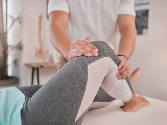
Medial knee pain after a direct blow or an awkward landing often points to injury in the structures that stabilize the inside of the knee. The valgus stress test is a hands-on exam used to assess stability, with particular attention to the medial collateral ligament (MCL).
During the test, the examiner applies a controlled valgus force to the knee and assesses pain, medial joint opening, and the quality of the ligamentous endpoint. Testing at different knee angles helps distinguish an isolated MCL injury from damage involving other stabilizing structures.
Where the knee gives way, at 30 degrees or in full extension, changes what the test is telling you. Here's how to perform it, read it, and know when it's pointing to more than an MCL sprain.
What is the valgus stress test?
The valgus stress test is a clinical technique used to assess the integrity of the knee's medial structures. It's commonly performed when an MCL sprain or tear is suspected.
The MCL runs along the inside of the knee, connecting the femur to the tibia. It resists valgus loading, which occurs when the knee moves inward relative to the hip and foot, and contributes to control of tibial rotation and other multiplanar movements.1
An MCL injury may occur after:
A direct blow to the outside of the knee
A planted foot combined with knee rotation
An awkward landing that causes the knee to collapse inward
A fall that places stress across the medial knee
A collision during sports or recreational activity
Common findings include medial knee pain, localized tenderness, swelling, bruising, and a sense that the knee may give way. How pronounced these symptoms are depends on the location and severity of the ligament injury.2
The valgus stress test places controlled tension on the MCL and related medial structures. The examiner watches for symptoms and compares medial joint movement with the opposite knee. Pain may suggest tissue irritation, while increased joint opening may indicate loss of ligament restraint.
The test is generally performed at two positions:
Approximately 20 to 30 degrees of knee flexion
Full or near-full knee extension
Each position tells you something different about what's contributing to knee stability.
How to perform the valgus stress test
Position the patient supine with the involved leg relaxed. Support the leg and place the knee at the desired testing angle.
For the valgus stress test at approximately 30 degrees:
Position the knee in about 20 to 30 degrees of flexion.
Stabilize the lateral side of the knee near the joint line.
Support the lower leg or ankle with the opposite hand.
Apply a controlled force that moves the lower leg laterally while directing the knee medially. Apply the force gradually.
Assess for pain, medial joint opening, and the quality of the endpoint.
Compare the response with the uninvolved knee.
Flexing the knee reduces the contribution of the joint capsule and several secondary stabilizers, which puts more emphasis on the superficial MCL as a restraint to valgus loading.3
Repeat the test with the knee in full or near-full extension, applying the same controlled valgus force while monitoring medial joint movement. In extension, several structures contribute to stability, including the MCL, joint capsule, cruciate ligaments, and posteromedial structures. Increased opening in this position may point to a broader injury rather than an isolated MCL lesion.1
A forceful or abrupt maneuver can increase discomfort, provoke muscle guarding, and make the result harder to read. The patient should stay as relaxed as possible so protective contraction doesn't mask joint motion.
What is a positive valgus stress test?
A valgus stress test is positive when it produces one or more of the following:
Familiar pain along the medial knee
More medial joint opening than the uninvolved side
A soft, delayed, or absent endpoint
Apprehension or protective muscle contraction
Symptoms that match the reported mechanism of injury
Pain and laxity give related but separate information. Pain without excess opening may occur with a mild sprain, tissue irritation, or partial injury where ligament continuity remains intact. Increased opening suggests the ligament is no longer providing its expected restraint.
A study of patients with traumatic knee injuries found that pain and laxity during valgus stress testing at 30 degrees were associated with MCL lesions identified on MRI. Diagnostic value improved when exam findings were combined with information about the injury mechanism.4
Interpreting valgus stress test findings
The knee angle at which instability shows up is one of the most useful parts of the exam.
Laxity at 30 degrees
Increased medial joint opening at approximately 30 degrees is consistent with MCL involvement. Since the superficial MCL carries much of the valgus load in this position, abnormal movement here may reflect partial or complete injury to that ligament.1
Document:
The amount of apparent joint opening
The presence and location of pain
The firmness of the endpoint
Differences between the involved and uninvolved knees
The patient’s confidence or apprehension during testing
Comparing with the opposite knee matters because normal ligamentous laxity varies from person to person. A small amount of symmetrical movement may be normal, while a clear side-to-side difference carries more clinical weight.
Laxity in full extension
Medial opening with the knee extended raises concern for injury beyond the superficial MCL. The posterior oblique ligament, posteromedial capsule, anterior cruciate ligament, posterior cruciate ligament, or other stabilizing tissues may be involved.1
Instability in both extension and flexion calls for a broader ligament exam, which may include the Lachman test, posterior drawer test, dial test, varus stress test, and assessment of rotational stability based on the suspected injury pattern.
MCL injury grades
MCL injuries are commonly described using three grades:
Grade I: A mild sprain involving microscopic fiber damage. Medial tenderness and pain may be present, but the valgus stress test typically shows no meaningful laxity and a firm endpoint.
Grade II: A partial ligament tear. The test may produce pain and increased medial movement, but a recognizable endpoint remains.
Grade III: A complete tear associated with marked medial laxity and a soft or absent endpoint. Grade III injuries may occur in isolation, though related ligament or capsular damage should be considered.5
Manual grading is partly subjective. Swelling, pain, guarding, examiner technique, and natural side-to-side differences can all affect the apparent amount of opening. Document findings carefully rather than relying on a grade label alone.
Accuracy and limitations of the valgus stress test
The valgus stress test provides useful clinical information, but it isn't a stand-alone diagnosis.
Research assessing traumatic knee injuries found that pain during valgus stress testing at 30 degrees had a reported sensitivity of 78 percent and specificity of 67 percent. When laxity was used as the positive finding, sensitivity rose to 91 percent, while specificity dropped to 49 percent.4 These numbers suggest that an absence of both pain and laxity can help lower suspicion for an MCL lesion, but a positive result on its own doesn't confirm the diagnosis.
Several factors can affect test accuracy.
Pain and muscle guarding
Acute injuries may cause enough pain that the patient contracts the hamstrings or surrounding muscles during testing. Guarding can limit medial joint opening and make an unstable knee appear more secure. It can also produce pain before meaningful stress reaches the ligament.6
Examiner position and force
Changes in hand placement, knee angle, tibial rotation, and applied force can produce different findings. Consistent positioning and comparison with the uninvolved side reduce some of this variation.
Related injuries
Medial knee pain may come from the medial meniscus, pes anserine region, bone injury, joint capsule, or other soft tissues. A positive valgus stress test may reflect stress across more than one structure.
The mechanism of injury can help guide the rest of the exam. A direct lateral impact may raise suspicion for an MCL injury, while valgus loading combined with rotation may raise concern for associated meniscal or cruciate ligament damage.
Role of imaging
Imaging may be appropriate when the exam suggests a high-grade tear, multiple ligament injury, fracture, persistent instability, or symptoms that don't match the initial findings.
MRI can show the location and extent of MCL damage and identify related injuries. Stress radiography can quantify medial joint opening under load, and dynamic ultrasound can provide real-time information about ligament behavior and medial joint gapping.7
Imaging findings should still be interpreted alongside symptoms, function, and clinical exam results.
How this looks in practice: A clinical example
A recreational soccer player reports medial knee pain after an opposing player struck the outside of the planted leg. The athlete felt the knee move inward but didn't hear a pop. Mild swelling developed later that day.
Examination reveals tenderness along the proximal MCL and pain during the valgus stress test at 30 degrees. There is a small increase in medial joint opening compared with the other knee, though a firm endpoint remains. Valgus testing in full extension doesn't reveal increased motion.
This pattern is consistent with a partial MCL injury. The knee should still be screened for meniscal and cruciate ligament involvement, and follow-up testing can track changes in pain, endpoint quality, side-to-side movement, and tolerance for functional tasks over time.
The value of the valgus stress test isn't a single yes-or-no answer. It's the pattern, where the knee opens, how much, and what stops it, that tells you whether you're looking at a sprain to manage or an injury to refer out.
Ready to enhance your orthopedic assessment skills?
Subscribe today to unlock full access to our comprehensive library of orthopedic special tests, including video demonstrations, clinical pearls, and expert-led guidance to support confident diagnosis and effective treatment planning.
To continue building your clinical expertise, visit our PT CEU page for in-depth courses, certifications, and guided learning paths.
References
Vosoughi F, Rezaei Dogahe R, Nuri A, Ayati Firoozabadi M, Mortazavi J. Medial collateral ligament injury of the knee: A review on current concept and management. Archives of Bone and Joint Surgery. 2021;9(3):255–262. https://pmc.ncbi.nlm.nih.gov/articles/PMC8221433/
American Academy of Orthopaedic Surgeons. Collateral ligament injuries. OrthoInfo. https://orthoinfo.aaos.org/en/diseases--conditions/collateral-ligament-injuries/
Aronson PA, Gieck JH, Hertel J, Rijke AM. Tibiofemoral joint positioning for the valgus stress test. Journal of Athletic Training. 2010;45(4):357–363. https://pmc.ncbi.nlm.nih.gov/articles/PMC2902029/
Kastelein M, Wagemakers HPA, Luijsterburg PAJ, et al. Assessing medial collateral ligament knee lesions in general practice. American Journal of Medicine and Sports. 2008;36(8):1486–1492. https://pubmed.ncbi.nlm.nih.gov/18954845/
Chen L, Kim PD, Ahmad CS, Levine WN. Medial collateral ligament injuries of the knee: Current treatment concepts. Current Reviews in Musculoskeletal Medicine. 2008;1(2):108–113. https://pmc.ncbi.nlm.nih.gov/articles/PMC2684213/
Manske RC, Prohaska D, Lucas B. Recent advances following anterior cruciate ligament reconstruction: Rehabilitation perspectives. Physical examination and imaging of acute multiple ligament knee injuries. North American Journal of Sports Physical Therapy. 2008;3(4):197–209. https://pmc.ncbi.nlm.nih.gov/articles/PMC2953340/
James EW, Williams BT, LaPrade RF. Stress radiography for the diagnosis of knee ligament injuries: A systematic review. Clinical Orthopaedics and Related Research. 2014;472(9):2644–2657. https://pmc.ncbi.nlm.nih.gov/articles/PMC4117881/
Meet the Author


Subscribe to Our Newsletter
Related Posts
April 21, 2025
McMurray Test: A Clinical Guide for Assessing Meniscal Injuries
By Medbridge

November 10, 2025
Lachman Test: Purpose, Procedure, and Clinical Relevance
By Medbridge

November 15, 2024
Top Exercises for the Early Stages of Knee Pain Recovery
By Medbridge

September 9, 2024
Top 10 Essential Strategies for ACL Rehabilitation Success
By Kyle Matsel PT and Phil Plisky