Relative Stiffness: What You Know, What You Don't, and Why It Matters
Unlock more effective assessments by understanding the layers behind mobility deficits. Explore how relative stiffness impacts patient movement—and how to address it through smarter physical exams and targeted interventions.
July 18, 2022
8 min. read
Performing a physical exam on a patient is a process of peeling back layers of a problem.
Does a particular movement relate to symptoms or dysfunction? Does your patient move this way by choice, by habit, or as compensation? What contributes to the trade-off? And then what contributes to that?
Although many may consider the strength-control-mobility paradigm the final layer, we can go deeper into the latter.
Mobility Deficits and Relative Stiffness
Mobility deficits break down into, effectively, two broad contributors: flexibility deficits and imbalances of relative stiffness. The latter, relative stiffness, presents objectively and behaves visually as a mobility deficit, but may relate to motor control deficiencies. It's easy to assess during a mobility assessment; however, the results greatly impact treatment direction.
What Is a Flexibility Deficit?
A true flexibility deficit is a lack of structural range of motion, due to either bony or connective tissue, or muscular limitations. In the case of limited muscle length, there is a decreased length and quantity of sarcomeres. This trait is both adaptive and trainable. Research shows that four-week immobilization periods can increase or decrease sarcomere length by 21 percent and 40 percent.
The term adaptive shortening refers to such muscle length deficits. Patients may display this after a period of casting, as when six weeks in an above-elbow cast leads to inadequate elbow extension, or due to lifestyle, such as when years in a seated job leads to inadequate true hamstring or psoas length. Individuals with a mobility-demanding history, such as former gymnasts or swimmers, may display the opposite trait—high amounts of a structural range of motion.
What Are Relative Stiffness and Relative Flexibility Imbalances?
As Sahrmann, Azevedo, and Van Dillen have described, relative stiffness refers to the passive tension (elasticity) and resting tone (on-ness) of muscles. Muscles in adjacent body regions may have differing passive tension levels due to size and collagen quantities, or different resting tones due to nervous system activity. These imbalances in relative stiffness can lead to a lopsided tug-of-war across joints at rest or when moving. This is referred to as relative flexibility.
Resting muscle tone, or active stiffness (the on-ness of a muscle at rest) responds quickly, but briefly. The above video uses resistance bands (black = very stiff, yellow = not stiff at all) to illustrate varying levels of muscle stiffness. A dynamic warm-up or a massage, for example, might temporarily make a very stiff muscle (imagine a black band) much less stiff (like a yellow band).
Now imagine two bands tied end-to-end with a knot in-between. As the adjacent muscles pull to actuate joints, imagine the bands pulling to move the knot. Picture how this might be different with two very stiff muscles (black bands that do not move very far); two muscles that are not stiff at all (yellow bands that do move very far); or two muscles whose stiffness levels differ (a yellow and a black band, pulling the knot off to one direction). As you read on, consider when these combinations may be ideal or suboptimal.
Assessing Relative Stiffness Imbalances
Relative stiffness imbalances appear in the same places one might find true flexibility deficits.
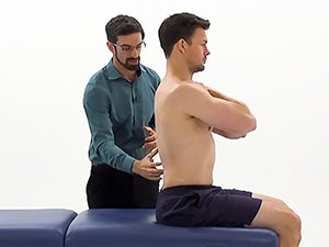
For the shoulder, the example above depicts an assessment of relative stiffness of the latissimus dorsi versus the abdominals. This patient has a full passive range of motion with the rib cage passively stabilized (the latissimus dorsi is a flexible yellow band, not a relatively stiff black band), and without rib cage stabilization (the abdominals and latissimus dorsi both are yellow bands, allowing full shoulder motion, with balanced relative stiffness). If his latissimus dorsi were a black band, his rib cage would elevate during end-range shoulder flexion, and his black band latissimus dorsi would win the tug-of-war with his yellow band abdominals.
This example with a bent-knee fallout looks at the relative stiffness of the lumbopelvic region—specifically, the obliques versus hip adductors and medial rotators. This patient has full hip range of motion but a relative stiffness imbalance between her hip adductors/medial rotators (black band) and her obliques (yellow band). Her falling knee pulls her pelvis toward it, inducing lumbar rotation. With the active engagement of her obliques, she turns her lumbopelvic yellow band into a black band.
In the above example, a seated knee extension is used to assess lumbopelvic relative stiffness. In the suboptimal movement demonstration, this patient's black band hamstrings pull his yellow band lumbar spine into flexion during seated knee extension. In the optimal demonstration, the patient actively engages his lumbar extensors, turning them into a black band to balance their relative stiffness against that of his hamstrings.
Implementation and Intervention
Now that we've explored the assessment of relative stiffness and imbalances across body regions, let's examine how to apply these concepts broadly and determine treatment direction.
Remember that what appears to be a flexibility deficit may be an issue of relative flexibility. To detect if a mobility deficit is a relative stiffness imbalance (black band tied to a yellow band), consider simulating the effect of turning the adjacent yellow band black.
For example, when performing a prone knee flexion test for hip flexor length, one might cue the patient to actively engage their trunk flexors and assess for a change in the test result. An increased apparent hip flexor length is a positive change and suggests that the hypothesized mobility deficit stems from a relative stiffness imbalance, a black band hip flexor tied to yellow band abdominals.
Remember also that patients may present with "yellow band-yellow band" or "black band-black band" combinations of relative stiffness across a joint or body region, and that this may be ideal (well-balanced) or suboptimal (balanced but too flexible or too stiff overall).
Now that you've assessed relative stiffness (muscle groups acting as black bands or yellow bands) and determined what has led to suboptimal body region movement, you will need to implement treatment. Let's look at how to turn a yellow band black or a black band yellow.
Turn a Yellow Band into a Black Band (Increase Relative Stiffness)
This example demonstrates a swimmer in whom the latissimus dorsi is a black band with greater relative stiffness than the abdominals, which act as a yellow band. When the shoulder is in full flexion, the yellow band gives way to the black band, and the rib cage flares. By engaging his abdominals (turning the yellow band black), he eliminates the relative stiffness imbalance with black band abdominals and black band latissimus dorsi, he can achieve full shoulder flexion without rib flare.
Beyond increasing active intra-muscular stiffness, isometric training has been demonstrated to be effective in improving passive stiffness and resting tone, in addition to helping achieve local pain inhibition. Further, the training required to increase passive stiffness and resting tone may improve regional motor control, with implications for patient self-efficacy and injury prevention.
Turn a Black Band Into a Yellow Band (Decrease Relative Stiffness)
In absence of significant movement or lifestyle changes, stretching has only short-lived effects on relative stiffness imbalances, lasting at least 90 minutes but returning to baseline by the next day.
In this video, the patient's latissimus dorsi is again a black band with greater relative stiffness than the abdominals. The clinician uses an inhibitory technique called post-isometric relaxation (PIR) to turn the black band yellow and decrease latissimus dorsi relative stiffness to correct the movement fault.
While the first example increased abdominal relative stiffness and turned a yellow band black, this example reduces latissimus dorsi relative stiffness, which turns the black band yellow. In both cases, the end result is a correction of the relative stiffness imbalance.
The Bigger Picture
If you've already begun using symptom modification procedures (SMPs) in your practice, you can smoothly integrate the assessment of relative stiffness. If not, we suggest you read Are Special Tests Necessary? Why You Should Use Symptom Modification Procedures Too.
Symptom modification procedures have three key benefits:
Improved patient buy-in via rapid demonstration of faults and corrections
Simplification of the physical exam
More effective selection of interventions
Watch the above video for a brief dive into SMPs, including how to apply passive and active correction or cueing to a patient's symptomatic movement. In assessing for relative stiffness imbalances, SMPs may include passive stabilization of yellow band regions, passive overpressure of black band regions to test for available range of motion, or cueing for active stabilization of yellow band regions.
Whether the findings are positive or negative, testing for relative stiffness imbalances as part of your SMPs enables you to quickly detect and address the true cause of the patient's key impairments, while helping the patient readily understand a complex concept.
Physical exams allow you to examine the layers of a patient's complaint or symptoms. When you encounter what appears to be a soft tissue mobility deficit, look deeper to investigate whether a relative flexibility deficit is masquerading as a muscle length restriction. You can test for it as part of your symptom modification procedures, and doing so will enable you to provide more targeted, informed, and efficacious treatment.
To learn more, Jared Vagy's series of courses on the movement system offer a deeper exploration into relative stiffness as well as other related concepts.
Meet the Authors


Subscribe to Our Newsletter
Related Posts

December 1, 2021
Ditch the Treatment Table: How to Assess and Treat with Reference and Relative Motion
By Jared Vagy and Joseph Derian

January 10, 2025
Top 5 Exercises for Hip Osteoarthritis
By Medbridge

July 6, 2023
Adapting Manual Therapy to Telehealth and Virtual Care
By Jared Vagy and Joseph Derian

August 28, 2023
Reaching Alignment: Joint Centration for Athletes
By Jared Vagy and Tyson Matsumoto