4 Ways to Feel Your Way Through Palpation of the Foot and Ankle
Plantar heel pain is a common condition, affecting around two million people in the United States each year1 and comprising 15% of all foot and ankle problems among people who seek professional care.2
A number of different structures contribute to cause heel pain.1 These structures are also loaded differently during movement. Therefore, identifying the specific site of pain during movement can reveal clues about how to help your patient.
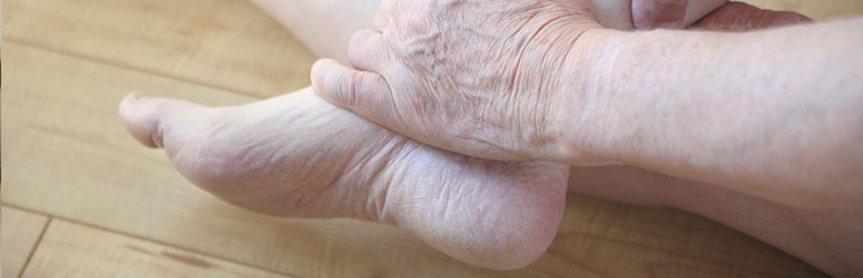
Palpation of the foot and ankle can be a critical tool to reveal the issue and assist in a positive patient outcome. Here are four tips to improve your palpation skills!
1. Practice
Trust in one’s hands requires practice. Palpation is a motor skill like any other we learned in physical therapy school. If we don’t practice regularly, we risk losing the ability to palpate accurately. To help focus your practice try the following:
- Schedule a reoccurring practice session with colleagues
- Keep a list of structures handy (and your favorite anatomical atlas)
- Make your practice sessions all about patients
There are not many studies that have characterized the reliability of palpation for the foot and ankle. The existing literature shows that physical examination tests and measures based on palpation can demonstrate reliability that ranges from good to excellent.3 Most structures are superficial and distinct enough that perhaps challenges to reliability observed for palpation elsewhere in the body, like the spine, could be less problematic in the foot and ankle.
2. Let the Patient Be Your Guide
The patient should guide the palpation and serve as your barometer. Before diving into a painful area, make sure the patient identifies it first. Ask your patient to “point to the painful area with one finger” to help prioritize the most important area to investigate.
As you palpate, make sure to keep your patient in charge to maintain trust. Build your manual pressure carefully so that you only use a level of pressure necessary to reproduce symptoms or assess tissue quality. Ask for feedback from your patient early and often throughout the palpation. Use one closed (yes/no) question at a time.
3. Have a Plan
Common structures you should be comfortable palpating in people with plantar heel pain include the plantar heel fat pad and plantar fascia (aponeurosis). Other structures often implicated in plantar heel pain involve medial structures of the foot and ankle, such as the tibial nerve (including medial branch of the calcanear nerve and medial plantar nerves), tibialis posterior tendon, and flexor digitorum longus.4
These potential origins of heel pain show the importance of planning your approach. A systematic approach to palpation can make your process efficient and inform your clinical reasoning process “on the fly,” to make treatment and prognosis decisions and to educate your patient.
4. Know Anatomy
Palpation involves the ability to mentally reconstruct the anatomy under your fingertips. A thorough knowledge of foot and ankle anatomy is necessary for accurate palpation.
As movement experts, we should know how the structures we palpate behave with loading. This kinesiological understanding can directly help with palpation. For example, the windlass test is a valuable test for identifying the plantar fascia.1 Dorsiflex the great toe and inspect the medial longitudinal arch for a band of tissue that becomes more prominent – that’s the plantar fascia (aponeurosis)! Can’t find the tibialis posterior tendon? Try to use the manual muscle test position to see whether the tendon becomes more prominent distal to the tip of the medial malleolus of the tibia.
While palpation is an important aspect of evaluating a patient with plantar heel pain, it’s just one part of combining information about pathoanatomy and differential diagnosis with clinical skills to optimize your patient’s outcome.
- Martin RR, Davenport TE, Reischl SF, McPoil TG, Matheson JW, Wukich DK, McDonough CM (2014). Heel pain-plantar fasciitis: revision 2014. Journal of Orthopaedic and Sports Physical Therapy. 44: 11: A1-33.
- Rome K, Howe T, Haslock I. Risk factors associated with the development of plantar heel pain in athletes. Foot. 2001; 11:119-125.
- Wrobel JS, Armstrong DG (2008). Reliability and validity of current physical examination techniques of the foot and ankle. Journal of the American Podiatric Medical Association; 98(3): 197-216.
- https://www.ncbi.nlm.nih.gov/pubmed/25361863